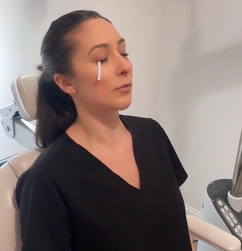
Dry Eye Exams
Dry eye exams are problem focused visits that consist of an array of tests including:
Schirmers Test
Measures tear production to rule out aqueous deficiency
Meibography
Captures an image of the meibomian glands surrounding the lids in order to evaluate for segmentation, thickening and atrophy.
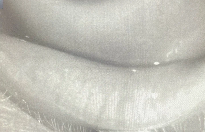
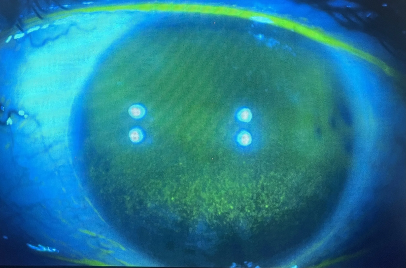
NaFL Staining
Fluorescein staining is used to evaluate your tear break up time and evaluates for corneal cell damage.
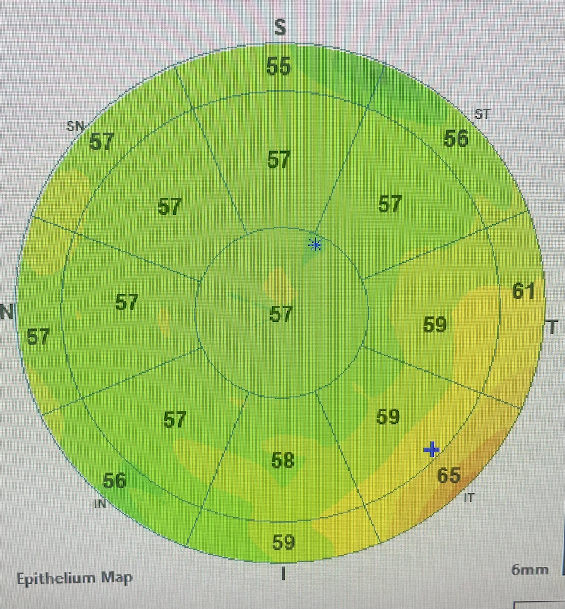
Epithelial Mapping
This advanced diagnostic OCT technology maps out the integrity of the corneal epithelium in order to monitor change over time and is useful for grading the severity of dry eye.
© 2026 Sorella Optique and Eyecare. All rights Reserved - Accessibility Statement - Privacy Policy - Sitemap
Managed and Designed by ![]()